Probe tip positioning into involved furcations of mandibular molars, poster #136
Authors
Guindy JS, Manolakis K, Pajarola J, Schupbach P, Zappa U. Department of Periodontology and Cariology, School of Dental Medicine, University of Basel, Basel, Switzerland
Date of Publication
J Dent Res 76 (IADR Abstracts) 1997
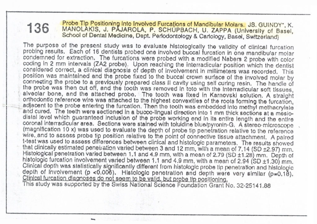
The purpose of the present study was to evaluate histologically the validity of clinical furcation probing results. Each of 16 dentists probed one involved buccal furcation in one mandibular molar condemned for extraction. Involved furcations were probed with a modified Nabers 2 probe with color coding in 2mm intervals (ZA2 probe). Upon reaching the interradicular position considered correct by the dentist, a clinical diagnosis of depth of involvement in millimeters was recorded. This position was maintained and the probe tip was fixed to the buccal crown surface of the involved molar by connecting the probe to a previously prepared class II cavity using self-curing resin. The probe handle was then separated, and the tooth was removed in toto, involving the interradicular soft tissues, alveolar bone and the attached probe in one unit. The tooth was fixed in Karnovski solution. A straight orthodontic reference wire was attached to the highest convexities of the roots forming the furcation, adjacent to the probe entering the furcation. Then the tooth was embedded into methylmethacrylate and cured. All teeth were sectioned in a bucco-lingual direction into 1mm thick sections at a mesiodistal level, which guaranteed inclusion of the probe working end in its entire length and the entire coronal interradicular area. Sections were stained with Toluidin blue. A stereo-microscope was used to evaluate the probe tip depth penetration relative to the reference wire and to assess probe tip position relative to the point of connective tissue attachment. Results showed that clinically estimated penetration varied between 3 and 12 mm, with a mean of 7,14 mm. Histological penetration varied between 1,1 and 4,9 mm. Depth of histologic furcation involvement varied between 1,1 and 4,9 mm. Clinical depth was statistically significantly different from histologic probe tip penetration and histologic depth of involvement. Histologic penetration and depth were very similar. Clinical furcation diagnosis do not seem to be valid, but probe tip positioning does. This study was supported by the Swiss National Science Foundation Grant No. 32-25141.88


